Home » Without Label » Abdominal Anatomy - Normal Abdominal Anatomy Medical Illustration : We're going to take apart a plastic anatomy model and see what we can find in the abdomen.
Abdominal Anatomy - Normal Abdominal Anatomy Medical Illustration : We're going to take apart a plastic anatomy model and see what we can find in the abdomen.
Abdominal Anatomy - Normal Abdominal Anatomy Medical Illustration : We're going to take apart a plastic anatomy model and see what we can find in the abdomen.. The abdominal aorta enters the abdomen through the diaphragm at the level of the twelfth thoracic vertebre and continues to just below the umbilical area, where it splits into the right and left common iliac arteries. The majority of these organs are encased in a protective membrane termed the peritoneum. A hernia will usually cause a distinct bulge where the tissue or organ pushes through the muscle wall. Much information can be gathered from simply watching the patient and looking at the abdomen. The normal anatomy or organs imaged in a standard abdominal examination is explained below.
The abdomen (colloquially called the belly, tummy, midriff or stomach) is the part of the body between the thorax (chest) and pelvis, in humans and in other vertebrates. This requires complete exposure of the region in question, which is accomplished as follows: The diaphragm marks the top of the abdomen and the horizontal line at the level of the top of the pelvis marks the bottom. The component of the urinary system, kidney and the ureter. The rectus abdominis connects to the xiphoid process, a bony landmark at the bottom of the sternum.
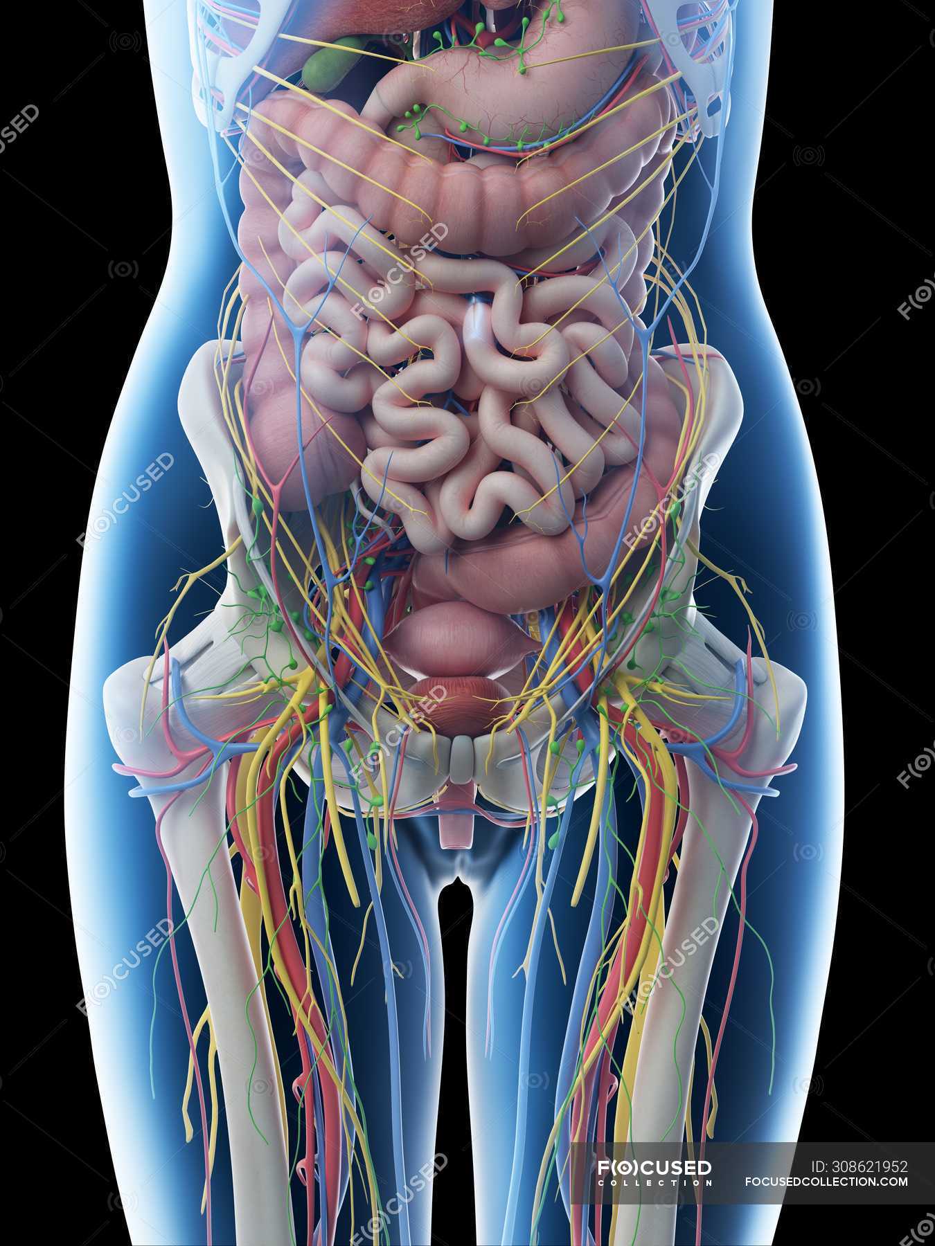
Female Abdominal Anatomy And Internal Organs Computer Illustration Medicine Digital Stock Photo 308621952 from st.focusedcollection.com The abdominal aorta enters the abdomen through the diaphragm at the level of the twelfth thoracic vertebre and continues to just below the umbilical area, where it splits into the right and left common iliac arteries. The abdominal cavity is the part of the body that houses the stomach, liver, pancreas, kidneys, gallbladder, spleen, and the large and small intestines. It also contains the spleen. The abdominal wall is defined cranially by the xiphoid process of the sternum and the costal margins, and caudally by the iliac and pubic bones of the pelvis. One of the easiest ways to tell if your pain is caused by a hernia or pulled stomach muscle is if you have a bulge or not. Stomach is a muscular bag forming the most distensible part of the human digestive system. It extends to the lumbar spine, which joins the thorax and pelvis and is a point of attachment for some abdominal wall structures 1 . Knowledge of abdominal wall anatomy is important to ensure safe placement of primary and secondary laparoscopic ports.
It is the long, flat muscle that extends vertically between the pubis and the fifth, sixth, and seventh ribs.
The abdominal wall is defined cranially by the xiphoid process of the sternum and the costal margins, and caudally by the iliac and pubic bones of the pelvis. Stomach is a muscular bag forming the most distensible part of the human digestive system. Definition (msh) that portion of the body that lies between the thorax and the pelvis. The region occupied by the abdomen is called the abdominal cavity, and is enclosed by the abdominal muscles at front and to the sides, and by part of the vertebral column at the back. The abdomen is the body region found between the thorax and the pelvis. Inferiorly the abdomen is open to the pelvis, communicating through the superior pelvic aperture (pelvic inlet). Abdomen anatomy the abdomen is comprised primarily of the digestive tract and other accessory organs which assist in digestion, the urinary system, spleen, and the abdominal muscles (shown below). Abdominal muscle strains don't cause a bulge or visible lump. Knowledge of abdominal wall anatomy is important to ensure safe placement of primary and secondary laparoscopic ports. Abdomen, in human anatomy, the body cavity lying between the chest or thorax above and the pelvis below and from the spine in the back to the wall of abdominal muscles in the front. Definition (nci) of, or related to, the abdomen. In anatomy and physiology, you'll learn how to divide the abdomen into nine different regions and four different quadrants. The abdomen (colloquially called the belly, tummy, midriff or stomach) is the part of the body between the thorax (chest) and pelvis, in humans and in other vertebrates.
This mri abdomen axial cross sectional anatomy tool is absolutely free to use. The component of the urinary system, kidney and the ureter. Skin, superficial fascia, muscles and associated fascia, and parietal peritoneum. The abdominal wall is defined cranially by the xiphoid process of the sternum and the costal margins, and caudally by the iliac and pubic bones of the pelvis. The abdominal wall surrounds the abdominal cavity, providing it with flexible coverage and protecting the internal organs from damage.
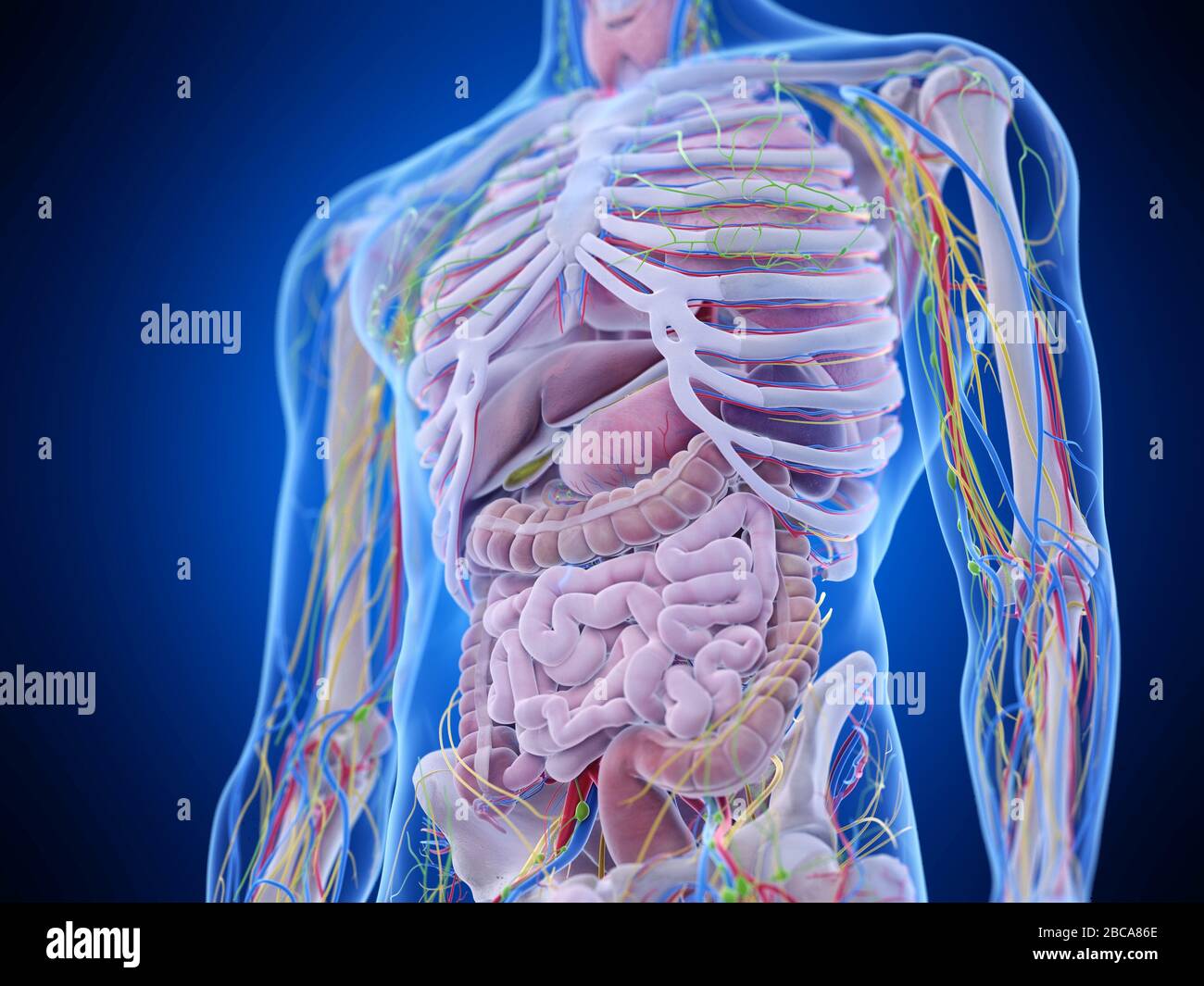
Abdominal Anatomy Illustration Stock Photo Alamy from c8.alamy.com The normal anatomy or organs imaged in a standard abdominal examination is explained below. Having to do with the abdomen, which is the part of the body between the chest and the hips that contains the pancreas, stomach, intestines, liver, gallbladder, and other organs. The area occupied by the abdomen is called the abdominal cavity. It is an artery, meaning that it carries blood away from the heart. We're going to take apart a plastic anatomy model and see what we can find in the abdomen. The abdomen is the body region found between the thorax and the pelvis. The rectus abdominis connects to the xiphoid process, a bony landmark at the bottom of the sternum. Topical anatomy of the abdomen.
Then liver & spleen) palpate 4 quadrants abdomen (superficial then deep) assess for kidney area pain (cvat) wash hands time target:
A hernia will usually cause a distinct bulge where the tissue or organ pushes through the muscle wall. It is the long, flat muscle that extends vertically between the pubis and the fifth, sixth, and seventh ribs. Having to do with the abdomen, which is the part of the body between the chest and the hips that contains the pancreas, stomach, intestines, liver, gallbladder, and other organs. We're going to take apart a plastic anatomy model and see what we can find in the abdomen. Definition (nci) of, or related to, the abdomen. The normal anatomy or organs imaged in a standard abdominal examination is explained below. Its superior aperture faces towards the thorax, enclosed by the diaphragm. The abdomen is the front part of the abdominal segment of the trunk. The abdominal wall is defined cranially by the xiphoid process of the sternum and the costal margins, and caudally by the iliac and pubic bones of the pelvis. The abdominal wall surrounds the abdominal cavity, providing it with flexible coverage and protecting the internal organs from damage. It is bounded superiorly by the xiphoid process and costal margins, posteriorly by the vertebral column and inferiorly by the pelvic bones and inguinal ligament. The abdomen contains all the digestive organs, including the stomach, small and large intestines, pancreas, liver, and gallbladder. The majority of these organs are encased in a protective membrane termed the peritoneum.
Then liver & spleen) palpate 4 quadrants abdomen (superficial then deep) assess for kidney area pain (cvat) wash hands time target: The rectus abdominis connects to the xiphoid process, a bony landmark at the bottom of the sternum. The component of the urinary system, kidney and the ureter. It is the long, flat muscle that extends vertically between the pubis and the fifth, sixth, and seventh ribs. To reach this goal and minimize complications, every reproductive surgeon requires a thorough knowledge of pelvic anatomy.
Abdominal Anatomy Radiology Students Of A M S Facebook from lookaside.fbsbx.com The abdominal wall surrounds the abdominal cavity, providing it with flexible coverage and protecting the internal organs from damage. Abdominal anatomy includes a major element of the gastrointestinal, system, the caudal end of the oesophagus, stomach, large and small intestine, liver, pancreas and the gallbladder. These organs are held together loosely by connecting tissues. Then liver & spleen) palpate 4 quadrants abdomen (superficial then deep) assess for kidney area pain (cvat) wash hands time target: The major organs of the abdomen include the small intestine, large intestine, and stomach. To reach this goal and minimize complications, every reproductive surgeon requires a thorough knowledge of pelvic anatomy. The abdominal aorta enters the abdomen through the diaphragm at the level of the twelfth thoracic vertebre and continues to just below the umbilical area, where it splits into the right and left common iliac arteries. Its superior aperture faces towards the thorax, enclosed by the diaphragm.
The abdomen is the front part of the abdominal segment of the trunk.
The abdominal wall surrounds the abdominal cavity, providing it with flexible coverage and protecting the internal organs from damage. A hernia will usually cause a distinct bulge where the tissue or organ pushes through the muscle wall. Knowledge of abdominal wall anatomy is important to ensure safe placement of primary and secondary laparoscopic ports. We're going to take apart a plastic anatomy model and see what we can find in the abdomen. Stomach is a muscular bag forming the most distensible part of the human digestive system. Abdomen, in human anatomy, the body cavity lying between the chest or thorax above and the pelvis below and from the spine in the back to the wall of abdominal muscles in the front. These two apertures, together with abdominal walls, bound the abdominal cavity. The area occupied by the abdomen is called the abdominal cavity. Abdominal anatomy includes a major element of the gastrointestinal, system, the caudal end of the oesophagus, stomach, large and small intestine, liver, pancreas and the gallbladder. To reach this goal and minimize complications, every reproductive surgeon requires a thorough knowledge of pelvic anatomy. The major organs of the abdomen include the small intestine, large intestine, and stomach. Use the mouse scroll wheel to move the images up and down alternatively use the tiny arrows (>>) on both side of the image to move the images.>>) on both side of the image to move the images. Having to do with the abdomen, which is the part of the body between the chest and the hips that contains the pancreas, stomach, intestines, liver, gallbladder, and other organs.