Home » Without Label » Pelvic Anatomy : Pelvic Anatomy / Anatomy the pelvis is a ring of bones located at the lower end of the trunk—between the spine and the legs.
Pelvic Anatomy : Pelvic Anatomy / Anatomy the pelvis is a ring of bones located at the lower end of the trunk—between the spine and the legs.
Pelvic Anatomy : Pelvic Anatomy / Anatomy the pelvis is a ring of bones located at the lower end of the trunk—between the spine and the legs.. The pelvis is the lower part of the torso. The pelvis is the lower portion of the trunk, located between the abdomen and the lower limbs. Use the mouse scroll wheel to move the images up and down alternatively use the tiny arrows (>>) on both side of the image to move the images.>>) on both side of the image to move the images. Anatomy of female pelvic area facebook twitter linkedin pinterest print fertility and reproductive health pelvic floor disorders fertility, pregnancy and childbirth women's health. Sacrum (the large triangular bone at the base of the spine)
Pathologic conditions of the pelvis may reach the abdomen and beyond; The pelvis is the lower part of the torso. When you are taking anatomy and physiology you will be required to know the anatomical structure locations of the pelvis. Laparoscopic anatomy of the female pelvic region. It is usually divided into two separate anatomic regions:
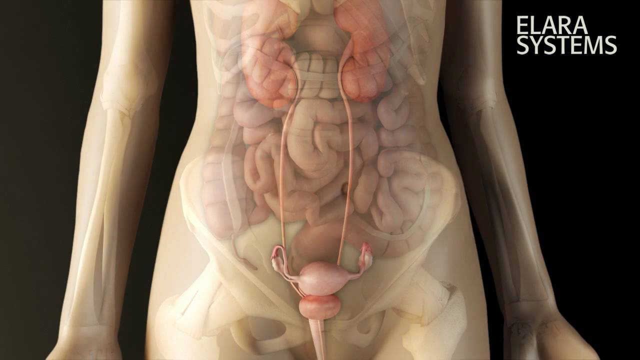
Female Pelvis Anatomy 3d Animation Youtube from i.ytimg.com Ebraheim's educational animated video describes the anatomy of the pelvis, the bony structures, ligaments, muscles, blood supply, and nerves.this video a. This cavity is located within the lesser part of the pelvis, beneath the pelvic brim. Anatomy the pelvis is a ring of bones located at the lower end of the trunk—between the spine and the legs. The lining of the uterus. • pelvis begins at the iliac crests and ends at the symphysis pubis. Consisting of the pelvic girdle and perineum, it supports the urinary and reproductive organs. Anatomy of female pelvic area facebook twitter linkedin pinterest print fertility and reproductive health pelvic floor disorders fertility, pregnancy and childbirth women's health. Complete coverage of both conventional and endoscopic surgeries helps you master the full spectrum of surgical procedures.
A pelvic ultrasound is a noninvasive diagnostic exam that produces images that are used to assess organs and structures within the female pelvis.
Access to the left ureter, left iliac vessels, and left ovarian vessels can be gained by sharply incising the peritoneal sidewall attachment of the sigmoid colon; Ebraheim's educational animated video describes the anatomy of the pelvis, the bony structures, ligaments, muscles, blood supply, and nerves.this video a. A pelvic ultrasound allows quick visualization of the female pelvic organs and structures including the uterus, cervix, vagina, fallopian tubes and ovaries. The pelvis is a basin shaped bony structure formed by the combination of two pelvic bones (hip bones or innominate bones) and the sacrum. Ultrasound uses a transducer that sends out. Ischial spine ischial tuberosity coccygeus: It is strengthened and supported by several joints and ligaments. Inferior end of sacrum lateral attachment: Surgical anatomy of the female pelvis by laparoscopy. The pelvis is the lower portion of the trunk, located between the abdomen and the lower limbs. 2 pelvic anatomy 2.1 sacral promontory. • divided into the true and false pelvis by the iliopectineal line. This mri female pelvis sagittal cross sectional anatomy title tool is absolutely free to use.
Ischial spine ischial tuberosity coccygeus: This area provides support for the intestines and also contains the bladder and reproductive organs. The pelvic bones include the: The sacral promontory in its literal sense is the summit of the pelvis. When you are taking anatomy and physiology you will be required to know the anatomical structure locations of the pelvis.
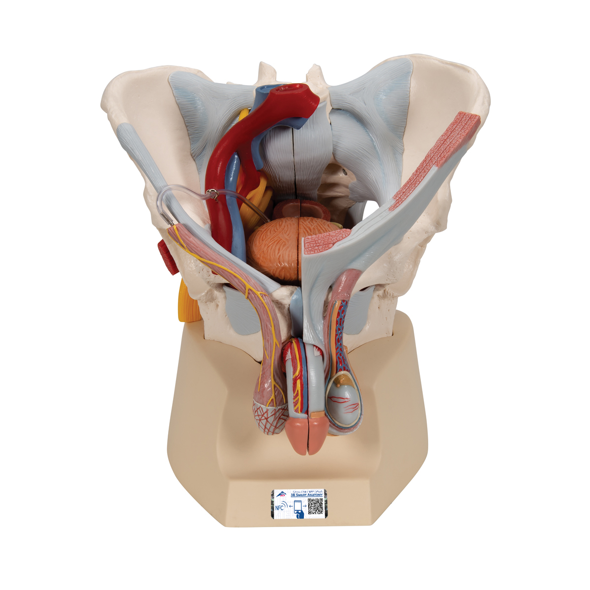
Male Pelvis Skeleton Model With Ligaments Vessels Nerves Pelvic Floor Muscles Organs 7 Part 3b Smart Anatomy 1013282 3b Scientific H21 3 Genital And Pelvis Models Anatomical Models from www.3bscientific.com Ultrasound uses a transducer that sends out. The sacral promontory in its literal sense is the summit of the pelvis. The lining of the uterus. The pelvic girdle (hip girdle) is formed by a single bone, the hip bone or coxal bone (coxal = hip), which serves as the attachment point for each lower limb. Sacrum (the large triangular bone at the base of the spine) Ischial spine ischial tuberosity coccygeus: Complete coverage of both conventional and endoscopic surgeries helps you master the full spectrum of surgical procedures. Use the mouse scroll wheel to move the images up and down alternatively use the tiny arrows (>>) on both side of the image to move the images.>>) on both side of the image to move the images.
The pelvis is inferior most part of the trunk.
However, their origin always lies at a level below the sacral promontory. Pelvic diaphragm, inferior view (gilroy et al.) atlas of anatomy 2nd ed., fig. 2 pelvic anatomy 2.1 sacral promontory. Ultrasound uses a transducer that sends out. This area provides support for the intestines and also contains the bladder and reproductive organs. It's located between the abdomen and the legs. • pelvis begins at the iliac crests and ends at the symphysis pubis. This cavity is located within the lesser part of the pelvis, beneath the pelvic brim. A pelvic ultrasound is a noninvasive diagnostic exam that produces images that are used to assess organs and structures within the female pelvis. This mri female pelvis sagittal cross sectional anatomy title tool is absolutely free to use. When you are taking anatomy and physiology you will be required to know the anatomical structure locations of the pelvis. The lining of the uterus. • divided into the true and false pelvis by the iliopectineal line.
It is usually divided into two separate anatomic regions: Ischial spine ischial tuberosity coccygeus: The right and left hip bones also converge anteriorly to attach to each other. Sacrum (the large triangular bone at the base of the spine) This quiz is unlabeled so it will test your knowledge on how to identify these structural locations (iliac crest, ischial spine, acetabulum, superior ramus of pubis, posterior superior/inferior iliac spine, lessier.
Anatomy Of The Pelvic Cavity Osmosis from d16qt3wv6xm098.cloudfront.net This area provides support for the intestines and also contains the bladder and reproductive organs. A pelvic ultrasound allows quick visualization of the female pelvic organs and structures including the uterus, cervix, vagina, fallopian tubes and ovaries. Inferior end of sacrum lateral attachment: Ebraheim's educational animated video describes the anatomy of the pelvis, the bony structures, ligaments, muscles, blood supply, and nerves.this video a. The male pelvis is different from a female's. Anatomy of female pelvic area facebook twitter linkedin pinterest print fertility and reproductive health pelvic floor disorders fertility, pregnancy and childbirth women's health. The lining of the uterus. It provides attachment to some important muscles in the region, and forms a cavity which accommodates several important internal organs.
Covering a compendium of gynecologic operations, including major and minor procedures and approaches, the techniques.
Surgical anatomy of the female pelvis by laparoscopy. • pelvis begins at the iliac crests and ends at the symphysis pubis. Pelvic surgery requires a comprehensive knowledge of the pelvic anatomy to safely attain access, maximize exposure, ensure hemostasis, and avoid injury to viscera, blood vessels, and nerves. When you are taking anatomy and physiology you will be required to know the anatomical structure locations of the pelvis. Anatomy of female pelvic area facebook twitter linkedin pinterest print fertility and reproductive health pelvic floor disorders fertility, pregnancy and childbirth women's health. Sacrum (the large triangular bone at the base of the spine) The pelvic region is the area between the trunk — or main body — and the lower extremities, or legs. Pelvis (hip) anatomy quiz for anatomy and physiology! This quiz is unlabeled so it will test your knowledge on how to identify these structural locations (iliac crest, ischial spine, acetabulum, superior ramus of pubis, posterior superior/inferior iliac spine, lessier. The pelvis is the lower part of the torso. A pelvic ultrasound allows quick visualization of the female pelvic organs and structures including the uterus, cervix, vagina, fallopian tubes and ovaries. It provides attachment to some important muscles in the region, and forms a cavity which accommodates several important internal organs. Access to the left ureter, left iliac vessels, and left ovarian vessels can be gained by sharply incising the peritoneal sidewall attachment of the sigmoid colon;